Dom Withers is Head of Radiotherapy Physics at Queen’s Hospital, Romford. Last year marked his 25th working full time in radiotherapy. Here he reflects on the changes in equipment, planning and the nature of his role over a quarter century.
This month marks my 25th anniversary of working full-time in radiotherapy. Back in 1999 I started as a Radiotherapy Physicist at Guy’s & St. Thomas’ Hospital in London. We had two ABB linear accelerators (linacs), a Theratron cobalt unit and a superficial caesium unit; and a Mobaltron cobalt unit and a Stabilipan orthovoltage unit. The latter two machines were pretty ancient. The Mobaltron had little handles you wound to check the positions; and it was rumoured that the Stabilipan was installed before the NHS was founded in 1948.
 None of the cobalt units or linacs had onboard imaging and no multi-leaf collimators to shape the treatment beam. Portal images could be taken by suspending a film in an envelope: this was then taken to a film processor in a dark room (usually by the most junior radiographer on set, or a student) and when developed it was then taken back to the set, displayed on a light box and image verification determined.
In 1999, most plans were created with a thick solder wire formed around the patient where they needed treatment. This was transferred to paper, stuck down and the outline drawn inside the wire using a pencil. The fluoroscopy unit was adjusted to the same position as the treatment beams, and X-ray films were taken with over-laying grids showing fields that defined the volume to be treated. The sizes of these fields were used to create the treatment volume on the paper, while the patient’s outline and treatment volume were then digitised into CadPlan, the treatment planning software. Beams were applied and adjusted to cover the treatment volume. The patient outline and dose distribution were printed out on A3 paper which was then signed by the doctor, and another sheet had all the treatment parameters for the beams that had to be typed into the linear accelerator system. The check of the dose calculation was done either by hand or by using an in-house MS-DOS program.
Captain Template and Princess Perspex
Some patients were having CT scans and treatment volumes could be drawn on each slice, moving away from cubic-shaped treatment volumes and allowing us to target the beam a little better while shielding some healthy tissue. For making treatment beam shapes other than oblongs, lead blocks had to be screwed to perspex templates that were mounted on the cobalt or linac collimator – one for each beam. Creating these was the first thing a junior physicist learnt to do in treatment planning: this earned the informal titles “Captain Template” for me and “Princess Perspex” for a colleague.
That’s quite a detailed description of what was done in 1999, but worth it to recall now as it’s astonishing to consider how radiotherapy technology has been transformed in the 25 years since. In my eight years at Guy’s & St. Thomas’ there were considerable developments. In 2000, replacement of the ABB linacs began and an Elekta linac was installed that had a multi-leaf collimator (MLC) that could shape the beam much better (and faster) than using lead blocks and templates. It also had portal imaging so we could see where we were treating quicker and easier. All very positive evolutions from the patient’s point of view, leading to quicker and more accurate treatments.
When the second ABB was replaced in 2001, we started to treat prostates with a higher treatment dose because we could see more clearly where we were treating. That meant we needed smaller margins around the prostate, and we could protect healthy tissue with MLCs from the full amount that they were getting from the “cube” shaped treatment volumes we did before.
Ever-changing technology
In 2002 we got a new treatment planning system, CMS, with a more sophisticated and more accurate dose calculation algorithm. In 2003 we started to do prostate brachytherapy using iodine seeds. Other developments included acquiring a simulator that could take single CT slices, replacement of the caesium and Stabilipan machines with kilovoltage units, installing software for recording and verifying linac treatments, electronic transfer of treatment plan parameters rather than manual copying, the creation of a multi-disciplinary team to implement intensity-modulated radiotherapy (IMRT), and replacement of the CT scanner. There was always change, always the chance to improve treatments, always the incentive to make things better. And speaking of change: Guy’s & St. Thomas’ was an early adopter of Agenda for Change (Outlining pay and conditions for NHS staff) and we were one of the few early adopter hospitals with a radiotherapy department.
In 2007 I attended a CMS treatment planning system users’ meeting in Madeira and started to pursue an idea I’d had to become their UK-based support operative. The creativity required and the fast pace of progress in treatment planning appealed to me (they still do) and I had learnt useful IT skills along the way: after an interview at the CMS European office in Freiburg in Germany, I was offered the job and started working from home as physics, applications and IT customer support for their treatment planning systems. Soon after I started, CMS was bought by Elekta. The job involved a lot of travel for training, demonstrations, conferences and installations. I visited 40 UK radiotherapy centres, and around another 40 in 25 countries in the region covered by the European office, including four in Africa and two in the Middle East; also many conferences in the UK and Europe. I got to see a huge range of practice.
Radiotherapy attracts innovators and those who want to do things better, and whilst my visits to hospitals were nominally to train other people, I always learnt something from each place and it was great to be able to share that combined wisdom and experience to other hospitals. The rest of the time I was at home covering support, learning about new versions or types of treatment planning programs, developing training materials, training customers in company offices, or attending conferences. IMRT became the new normal during this time, along with VMAT – Volumetric Modulated Arc Radiotherapy – where the gantry moved around whilst MLCs shaped the beam.
Queen’s Hospital in Romford
After several years the travel to train people in other hospitals that I had once loved, became too difficult with a young family at home. I was also yearning to be back in a clinical environment rather than a commercial one. In December 2015 the job of Head of Treatment Planning at Queen’s Hospital in Romford was advertised. It was a perfect fit: the right topic, at the right level, and geographically quite close. I worked out that the best way to get there from home was by motorbike, except that I had never ridden one before. I applied for a licence and began riding a motorbike at the age of 46 (a classic mid-life crisis perhaps?) to work at Queen’s Hospital. Queen’s had three Varian linacs, a CT scanner, and a kilovoltage superficial unit.
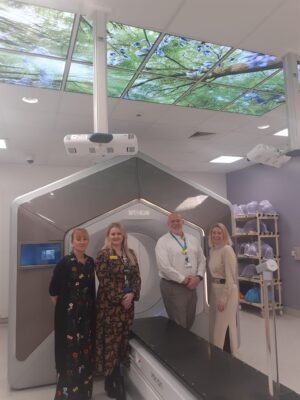
Going back to the clinic after eight years in industry was definitely a challenge, but I had great support from the head of department and others, and within six months I felt right at home. I had planning staff to manage, a new planning system to learn, a type of linac I hadn’t used before (Queen’s was all-Varian) and, of course, re-immersion into NHS working practices, administration and bureaucracy. Within 18 months of arrival, it was time to decide on the replacements of the department’s three linacs: this led to a period of rapid change and development in the department that does not seem to have stopped since. The decision was made in early 2017 at a higher level – and it was a bold decision: two of the three new machines would be Halcyons, a new ring-based linac whose existence had not even been made public by Varian. We became the third hospital in the world – second in Europe, first in the UK – to install and use a Halcyon linac in October 2017.
At the time Halcyon had only MV imaging and just one of its two banks of stacked MLCs being used for optimisation. Eight months later it became the first Halcyon version 2 in clinical use, with new kV-CBCT imaging and fully optimising MLCs. A second Halcyon was installed in 2018, and in 2020 – during Covid – it was upgraded to Ethos, capable of producing new treatment plans based on the CBCT taken at each treatment, taking into account changes in the patient shape from day to day. Stereotactic radiotherapy was rolled out nationally at this time too – it was a challenging few years, learning new techniques, with new technology, in a time of constant change. And that change has continued: upgrades to hardware, software, installation of surface guided systems, moving to a cloud-based oncology management system… it never stops.
And there was more change for me too. In early 2022 I became head of department, which has brought management-related challenges alongside the technical physics ones and more responsibilities. Although the Agenda for Change increment system has changed, I saw its wisdom in starting this role: you get the full pay for your band with the experience, and you better make sure you’re learning the whole time.
Three lessons from 25 years in radiotherapy
So what have been the biggest changes in 25 years of radiotherapy? I’ll limit it to three.
First – software. The hardware of mega-voltage radiotherapy equipment has barely changed in 25 years. Yes, there has been re-packaging – Tomotherapy, CyberKnife, Halcyon – but the physics and engineering is pretty much the same. One interesting change was actually a removal – taking out the filter that makes the “pointy” beam of a linac flat, which became a more obvious thing to do when IMRT and VMAT dominated: why flatten a beam that you are then going to make a different shape? The increase in rate at which energy came out of the machine, without a large chunk of metal in the beam, was a bonus.
But software improvements, taking advantage of speed improvements in computers, have allowed more complex plans to be produced, more sophisticated control of treatment machines to create IMRT and VMAT plans, and large oncology management systems that can receive, process and create the data needed for radiotherapy patients. Now AI-based contouring of anatomical structures on CT, MR and CBCT is common-place.
Second – imaging. The requirement for that student radiographer run down a corridor to develop a film feels so primitive now. First an imaging panel opposite the linac head taking planar images with the mega-voltage treatment beam, then kV generators and imaging panels creating planar images, then cone-beam CT images for MV and kV – these have transformed patient set-up accuracy and confidence in delivery. The development of linacs using MR technology for imaging can be included in this. Of course, developments in diagnostic PET, MR and CT have contributed to how radiotherapy plans are created as well.
Combing these two improvements, faster computing power and improved software can now produce treatment plans on CBCT or MR images taken immediately before treatment.
Third is less technology oriented – physicists are much closer to the patient than they were when I started. There was more of a distance, sometimes a proud distance, from direct patient involvement, and perhaps a more academic approach by physicists in a hospital environment. We were very much “behind the scenes” and highly technical, rather than clinical. We would work on the machines when there were no patients; we would wave goodbye to a treatment plan, once produced, never see it again, and move on to the next one. For a physicist now, involvement in techniques and technologies that take into account all aspects of the patient’s treatment is not limited to acceptance and commissioning, but we are more involved in the whole pathway from CT to final treatment – for example in motion management at CT such as breath-hold or 4DCT, imaging during treatment, evaluating the changes in plan delivery when analysing that imaging, surface-guided systems. We are coming out from behind the scenes a lot more, and this gives the modern radiotherapy physicist a deeper understanding of what patients go through – this can only be a good thing, as we try to develop techniques that are not just technically good but are appropriate to the real-life experience of patients.
The future of radiotherapy
What’s next? Cancer diagnosis rates are always increasing with better detection and an ageing population. In radiotherapy we have to play catch-up and treat more patients with, most likely, not the corresponding proportional increase in resources. Software and imaging will continue to improve, offering faster and more accurate ways to calculate treatment plans; improving imaging quality during treatment is a main goal for radiotherapy manufacturers to give better options for adapting treatment in real-time; more treatments will move to fewer fractions for the same effect – better for the patient, better patient throughput at the linacs; analysis of radiotherapy “big data” will hopefully be used to determine what works best.
To sum up – it’s never been boring, and the pace of change is unlikely to slow down. But thriving on the challenges of change is essential for ensuring we can continue to offer the best treatments for patients. The change is so relentless that at times I crave just a little boredom: perhaps that will have to wait until retirement!